
The printout slides out of the analyser while the patient is still being settled, and suddenly there are nine or ten numbers competing for your attention: pH, pCO2, pO2, bicarbonate, base excess, lactate, potassium, glucose. Under time pressure it is easy to stare at the sheet, fixate on the one value that looks frightening, and lose the thread. The reassuring part is that knowing how to interpret blood gas results is not about clever pattern spotting. It is about running the same short order every single time, so the analysis happens the same way whether you are calm or the resus bay is full.
This guide gives you that repeatable order, then covers the sample-type caveats that quietly change the numbers before anyone interprets them.
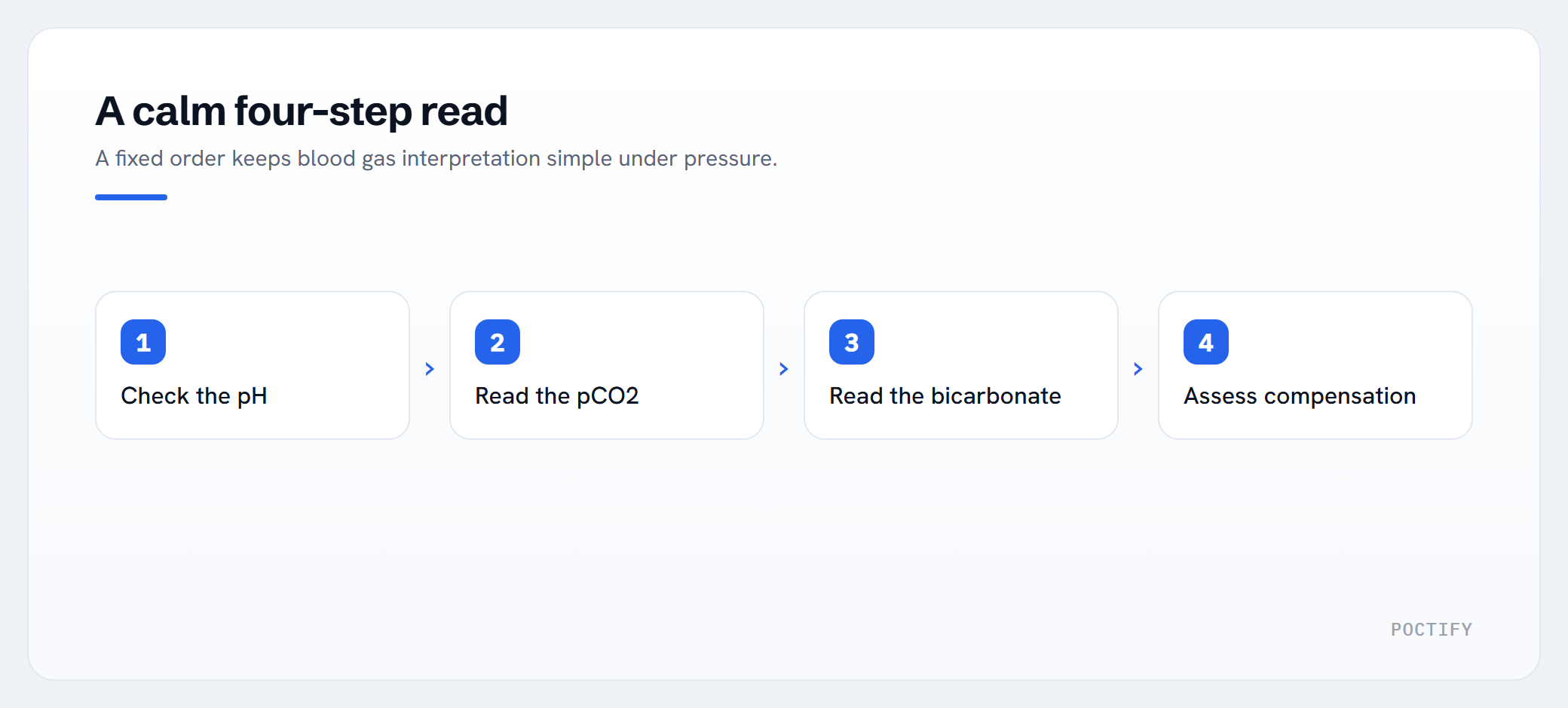
How to interpret blood gas results: the same four steps, every time
The aim of the first pass is narrow: classify the acid-base picture. You are not reaching a diagnosis, you are working out which of four primary problems you are looking at and whether the body is compensating.
Step 1: Read the pH first, and nothing else
Look only at the pH before any other value.
- Normal: 7.35 to 7.45
- Acidaemia: below 7.35
- Alkalaemia: above 7.45
This single number gives you the direction of the disturbance. Even when compensation has pulled the pH back toward normal, it usually sits on one side of 7.40, and that side tells you which process is winning. Commit to acid or alkali before you look any further.
Step 2: Read the respiratory picture (pCO2)
Carbon dioxide behaves as an acid gas: more CO2 means more acid and a lower pH.
- Normal pCO2: 4.7 to 6.0 kPa (35 to 45 mmHg)
- A high pCO2 pushes the pH down (respiratory acidosis)
- A low pCO2 pushes the pH up (respiratory alkalosis)
Now ask one question: does the pCO2 explain the pH? If the pH is low and the pCO2 is high, the lungs are a plausible cause.
Step 3: Read the metabolic picture (bicarbonate and base excess)
Bicarbonate is the main buffer base, so it moves the opposite way to acid.
- Normal bicarbonate: 22 to 26 mmol/L
- Normal base excess: -2 to +2 mmol/L
- A low bicarbonate, or a base excess more negative than -2, points to metabolic acidosis
- A high bicarbonate, or a base excess above +2, points to metabolic alkalosis
Base excess is simply a second way of reading the metabolic component, useful when the bicarbonate is borderline.
Step 4: Name the primary problem, then check for compensation
Match the value that moves in the same direction as the pH abnormality. That is your primary disorder.
| pH | pCO2 high | pCO2 low | HCO3 low | HCO3 high | |—-|———–|———-|———-|———–| | Low (acidaemia) | Respiratory acidosis | | Metabolic acidosis | | | High (alkalaemia) | | Respiratory alkalosis | | Metabolic alkalosis |
Then look at the other system. Compensation means the second system is shifting to drag the pH back toward 7.40: the kidneys retain bicarbonate over hours to days for a respiratory acidosis, and the lungs blow off CO2 within minutes for a metabolic acidosis. Two rules keep you out of trouble:
- In a single, compensated disorder the pCO2 and bicarbonate move in the same direction. If one is high and the other is low, suspect a mixed disorder.
- Compensation rarely returns the pH fully to normal. A pH that has overshot back past 7.40 suggests a second primary process, not tidy compensation.
A worked example of the method (numbers only)
Take a printout reading pH 7.30, pCO2 3.4 kPa, bicarbonate 13 mmol/L, base excess -10, lactate 6 mmol/L.
- Step 1: pH 7.30 is acidaemia.
- Step 2: the pCO2 is low, which on its own would raise the pH, so the lungs are not the cause. They are helping.
- Step 3: bicarbonate and base excess are both low, which fits metabolic acidosis, and that matches the pH.
- Step 4: a primary metabolic acidosis with respiratory compensation. The raised lactate points toward a high anion gap.
The method has classified the disturbance in seconds and without guesswork. This is classification only. The cause and the management belong to the bedside team and your local protocols.
Don’t skip the anion gap
When you find a metabolic acidosis, calculate the anion gap: sodium minus the sum of chloride and bicarbonate, normally around 8 to 12 mmol/L. A raised gap points to added acid such as lactate, ketones or certain toxins. A normal gap points to bicarbonate loss, for example from diarrhoea or renal tubular causes. Most gas analysers also report lactate at the same time, which often explains a raised gap immediately. If lactate is unfamiliar territory, the POCTIFY lactate explainer sets out what the result represents and the common reasons it rises.
The sample changes the number before you interpret it
Here is the part that rarely reaches the teaching whiteboard. A perfectly logical interpretation built on a flawed sample is still wrong. Three sample-type issues distort gas results before anyone applies the four steps.
Arterial versus venous: pO2 is the trap
A peripheral venous blood gas is quicker and kinder to obtain, and for acid-base work it is often good enough. A 2014 systematic review and meta-analysis in Respirology found that venous pH runs only about 0.03 below arterial with acceptable agreement, and venous bicarbonate agrees well too. The weak point is pCO2: the venous value carries wide limits of agreement and cannot be treated as an exact substitute for the arterial figure. A 2010 review in Emergency Medicine Australasia reached a similar position, noting that a normal venous pCO2 makes significant arterial CO2 retention unlikely and works as a screen, while an abnormal value should prompt an arterial sample.
The one number that does not transfer at all is pO2. Venous pO2 reflects how much oxygen the tissues have already extracted, not how well the lungs are oxygenating blood. You cannot assess oxygenation from a venous gas. Use pulse oximetry, and take an arterial sample when oxygenation genuinely matters.
Capillary blood gas has a defined place
Capillary sampling is not a poor relation. It has a real role, particularly in neonates and small children where arterial access is difficult and may be needed repeatedly. A properly arterialised sample, taken from a warmed, well-perfused heel or earlobe, gives a reasonable read on pH and pCO2. The caveats are firm: capillary pO2 is unreliable, squeezing the site adds venous and tissue fluid and provokes haemolysis, and air contamination shifts the result. Treat capillary oxygen values with caution and lean on saturations instead.
Occult haemolysis: the silent potassium error
Haemolysis releases potassium out of red cells, so a haemolysed sample can report a potassium far higher than the patient’s true level. With a gas syringe this is genuinely hazardous, because you cannot inspect the sample for the pink tinge of haemolysis the way a laboratory can with spun serum. The error is occult, and a falsely high potassium can trigger treatment a patient never needed. A difficult draw, a fine needle, excessive suction, an underfilled syringe or a long delay before analysis all raise the risk. If a potassium result does not fit the clinical picture, repeat it before acting. The POCTIFY potassium explainer covers why pseudohyperkalaemia happens and how sampling drives it.
A quick pre-analytical checklist
Before you trust any gas, confirm the basics:
- No visible air bubbles: bubbles raise pO2 and lower pCO2.
- Analysed promptly: a sample left at room temperature keeps metabolising, so pH and oxygen fall while CO2 and lactate climb. Analyse within about 15 minutes or chill the sample.
- Correct fill and the right syringe: too much liquid heparin dilutes the sample and lowers pCO2, bicarbonate and ionised calcium.
- Sampled cleanly: a traumatic draw is the usual route to occult haemolysis.
Make the numbers trustworthy at the point of care
A structured read only protects the patient if the result that reaches you is sound. That depends on the unglamorous layer around the analyser: in-date quality control, operators whose competency is current, and a clear record of which device produced which result. POCTIFY provides digital solutions for point-of-care testing, tailored to each clinic’s needs, and works with the devices and systems you already use to keep that pre-analytical and quality-control picture visible rather than assumed.
This article is educational and operational only. It does not provide medical advice, diagnosis or treatment guidance for any individual patient. Always follow your local clinical protocols and seek senior support when you need it.
If you want that quality-control and operator picture set up around your own analysers, talk to POCTIFY about tailored support for your service.
Frequently asked questions
What is the first thing to read on a blood gas?
Read the pH on its own before any other value. It tells you whether the overall picture is acidaemia (below 7.35), alkalaemia (above 7.45) or normal (7.35 to 7.45), and that direction anchors every step that follows.
Can a venous blood gas replace an arterial one?
For acid-base work it is often close enough. A 2014 Respirology meta-analysis found venous pH and bicarbonate agree well with arterial, while venous pCO2 has wider limits of agreement and works best as a screen. Venous pO2 cannot assess oxygenation at all, so use oximetry or an arterial sample when oxygenation matters.
When is a capillary blood gas appropriate?
It has a defined role, especially in neonates and small children where arterial access is difficult or repeated. A warmed, well-perfused, properly arterialised earlobe or heel sample gives a reasonable read on pH and pCO2. Capillary pO2 is unreliable, so treat oxygen values with caution and lean on saturations.
Why might potassium look falsely high on a blood gas?
Haemolysis releases potassium from red cells, so a damaged sample can report a level far above the patient’s true value. In a gas syringe this is occult because you cannot see the haemolysis. If a high potassium does not fit the clinical picture, repeat the sample before acting on it.
How can I quickly tell metabolic from respiratory acidosis?
After confirming acidaemia, check which value moves in the same direction as the low pH. A high pCO2 points to a respiratory cause, while a low bicarbonate or negative base excess points to a metabolic cause. If both pCO2 and bicarbonate move together you are likely seeing compensation, and if they move in opposite directions, suspect a mixed disorder.
How soon should a blood gas be analysed?
Within about 15 minutes, or chill the sample if there will be a delay. Cells keep metabolising in the syringe, which lowers pH and oxygen while raising CO2 and lactate, so a late sample can read meaningfully differently from the patient’s actual state.